MUT-IOE (Warsaw, Poland)
Institute of Optoelectronics (IOE), Military University of Technology (MUT)
The Institute of Optoelectronics at the Military University of Technology is a leading research institution in Poland dedicated to the development and application of lasers. Specific research areas in this field include: laser optics and electronics, laser systems, laser-matter interactions, laser distance measurement and detection, laser-generated secondary radiation sources, and application of lasers in nanotechnology and biomedicine.
The research team participating in the Lasers4EU project is the Laser Matter Interaction Group (LMI Group), which specializes in the development of laser-driven sources of X-rays and extreme ultraviolet (EUV) and their application in scientific research and modern technology.
Website: Laser Matter Interaction Group

Research highlights
 Secondary Sources
Development of laser-plasma sources of soft X-rays and extreme ultraviolet (EUV) based on a gas puff target [Opt. Express 30, 47867 (2022); Opt. Express 29, 20514 (2021); Phys. Plasmas 27, 073102 (2020)]
Plasma Physics
Generation of high-density cold plasmas by phtoionization of gases with intense EUV pulses [Phys. Plasmas 32, 023503 (2025); Phys. Plasmas 30, 103301 (2023); Phys. Plasmas 29, 093302 (2022)].
Cellular and Molecular Biology
Soft X-ray nanoimaging of cell structures [Appl. Sci. 2022, 12, 7030; Appl. Surf. Sci. 606 (2022) 154779; Int. J. Mol. Sci. 22, 7279 (2021)]
Imaging & Diagnostic Techniques
Development of new techniques for nanoimaging and spectroscopy studies [Opt. Express 30, 13491 (2022); J. Phys. & Chem. of Solids 164 (2022) 110623; Materials 2021, 14, 7337; Radiat. Phys. Chem. 175, 108086 (2020)].
Laser processing of materials
Surface modification of biomaterials with EUV pulses and EUV-induced cold plasma [J. Mater. Sci. 59, 11937 (2024); Appl. Surf. Sci. 606, 154779 (2022); Int. J. Mol. Sci. 21, 9679 (2020)]
Radiobiology and radiation chemistry
DNA damage by soft X-rays in the water window [Sci. Rep. 14, 28515 (2024)
Expertise
The LMI Lab has developed a range of experimental systems and workstations for research work conducted using soft X-rays and extreme ultraviolet (EUV). The systems and workstations are based on compact sources of this radiation with a laser-irradiated gas puff target. These devices have been applied in various fields of science and technology, including soft X-ray and EUV microscopy and tomography, soft X-ray absorption spectroscopy (NEXAFS and EXAFS), processing materials with EUV photons, soft X-ray and EUV radiation damage, modification of biomaterials, EUV photoionized plasma studies, metrology of soft X-ray and EUV optical elements and detectors, and others. The experimental setups and workstations are available for the external users and project partners for joint short-term experiments.
Equipment offered to external users
The LMI laboratory offers users various laser plasma sources of soft X-rays and extreme ultraviolet (EUV), a soft X-ray transmission microscope, an EUV transmission microscope, a soft X-ray contact microscope, a laboratory system for the X-ray absorption fine structure (XASF) spectroscopy in the soft X-ray range and a workstation for processing material using intense EUV pulses.
Fig. 1. High-energy Nd:YAG laser system. Credit: IOE
Laser plasma sources of soft X-rays and extreme ultraviolet (EUV)
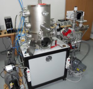
Laser plasma sources of soft X-ray and extreme UV radiation using a double-stream gas puff target are available to users for conducting studies of both the emission of radiation for its optimization and the application of the generated radiation. The targets are produced using a solenoid valve system consisting of two separate valves equipped with a special dual nozzle in the form of a circular hole surrounded by a hole in the form of a ring. The use of a double-stream gas puff target ensures high efficiency of X-ray and EUV emission without producing target debris. For target exposure, Lab LMI has several Nd:YAG laser systems generating laser pulses with a duration from 1 ns to 10 ns, and energy from 0.5 J to 10 J with repetition up to 10 Hz. Figure 1 shows the Nd:YAG Laser system generating laser pulses with energy up to 10 J.
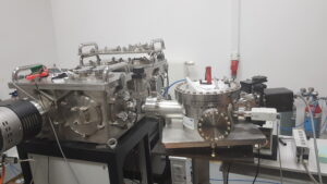
Fig. 2. Experimental station with the McPherson EUV spectrometer. Credit: IOE.
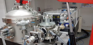
Fig. 3. Soft X-ray transmission microscope based on Fresnel and grazing incidence optics. Credit: IOE.
Soft X-ray transmission microscope
The soft X-ray transmission microscope (see Fig. 3) is based on a Fresnel zone plate as a microscope objective and an axisymmetric ellipsoidal mirror as a collector. The microscope uses radiation with a wavelength of 2.8 nm, generated in a laser plasma containing helium-like nitrogen ions. Using this microscope, images of both biological samples and solid nanostructures were obtained with a spatial resolution of 60 nm. The X-ray microscope is also equipped with a sample mounting system that allows it to be rotated around an axis perpendicular to the optical axis of the microscope, which allows for 2D images of the tested sample collected over the course of a rotation to reconstruct and subsequently display a 3D image.
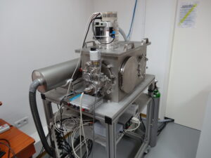
Fig. 4. EUV transmission microscope based on Fresnel and multilayer optics. Credit: IOE.
EUV transmission microscope
The EUV transmission microscope (see Fig. 4) uses radiation at a wavelength of 13.8 nm, which is obtained by means of an ellipsoidal mirror collector plated with Mo/Si layers, which selects this radiation from the broad spectrum of the argon laser plasma radiation. The EUV microscope was used to obtain images of solid nanostructures (nanowires and nanocrystals) with a spatial resolution of about 50 nm.
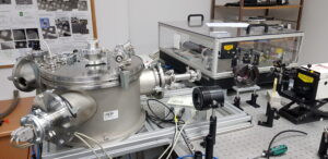
Fig. 5. Soft X-ray contact microscope. Credit: IOE.
Soft X-ray contact microscope
The soft X-ray contact microscope (see Fig. 5) also uses radiation from laser-produced argon plasma, but in the entire spectral range of the “water window”. The microscope has been used to obtain images of biological samples (cancer cells and stem cells) with a spatial resolution below 80 nm.
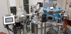
Fig. 6. Laboratory setup for XAFS spectroscopy. Credit: IOE.
Laboratory setup for XAFS spectroscopy
The laboratory system for the X-ray absorption fine structure (XASF) spectroscopy (see Fig. 6) in based on a laser plasma source with a krypton/xenon gas puff target producing soft X-ray radiation the wavelength range from about 1 nm to 10 nm. It allows measurements of XAFS spectra near the K-edges of low-Z elements (carbon, oxygen, nitrogen) and near the L-edges of medium-Z elements (transition metals). XAFS spectra are recorded using a reflection grating spectrograph equipped with a 2400 lines/mm grating and a CMOS camera. The recording of a XAFS spectrum in a single shot was demonstrated. The possibilities of XAFS measurements for gaseous or liquid samples and measurements in the reflection mode have been studied.
Workstation for EUV processing materials
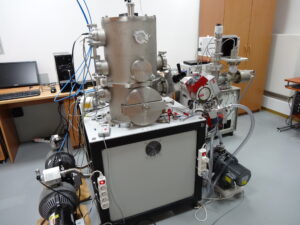
Fig. 7. Workstation for processing materials with EUV pulses and EUV-induced cold plasma. Credit: IOE.